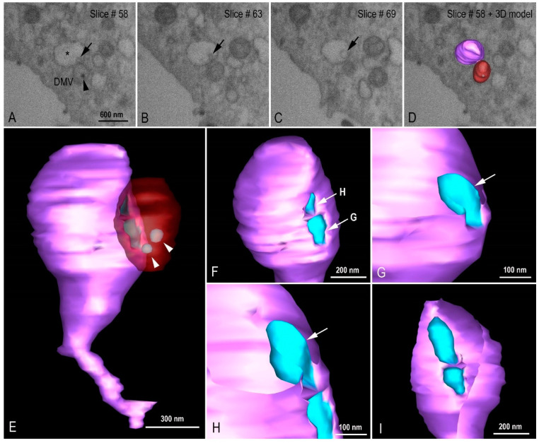
Figure 10.
Virion budding events at intermediate vesicle (IV). (A–D) FIB-SEM image sequence exhibiting different virus budding stages (arrows) at IV (asterisk) and the proximity with a double membrane vesicle (DMV). Virus particles can be observed inside the DMV (arrow head). (E–I) 3D model showing the DMV (red with transparence), virus particles (light blue, arrow heads), and the IV (purple). (F) Extra vesicular view of two virus particle budding regions. The arrows indicate the cutting plane showed in G and H. (G) Initial stage of virus particle budding. (H) Final stage of virus particle budding. (I) Intra vesicular view of the virus budding. See video in Supplementary Material (Video S2).

