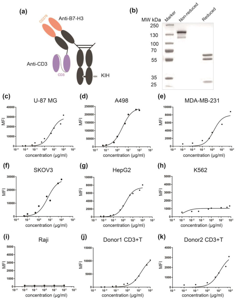
Figure 3.
Generation and characterization of αB7-H3/CD3 bispecific antibody. (a) An illustrative representation of the αB7-H3/CD3 format. The format comprised an anti-CD3 scFv fused to light chain of a monovalent anti B7-H3 via a (G4S)3 linker; (b) Coomassie blue-stained SDS-PAGE analysis of purified αB7-H3/CD3 containing three chains with the following molecular weights: 57, 55, and 28 kDa; (c–k) αB7-H3/CD3 bispecific antibody dose-dependently binds to B7-H3+ tumor cells and CD3+ T cells, evaluated using flow cytometry. MFI values (control/maximum concentration of 10-2#c) of these cell lines were, respectively, 205/31,800, 7500/226,444, 480/5700, 350/27,950, 150/8065, 480/1550, 125/155, 50/10,300, and 195/3100.

