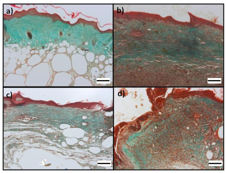
Figure 8.
Photomicrographs of wound at day 14: Masson’s trichrome for evaluation of collagen in histological sections of excisional wound site obtained from a diabetic wound: (a,b) images from different mice treated Plg-loaded fibrin scaffold show a qualitative greater collagen deposition compared to (c,d) images from different mice treated with Plg-unloaded fibrin scaffold. Collagen fibers stained with light green SF yellowish appear green. All figures show epithelialization and granulation tissue based on the state of wound healing; OM 100× (scale bare = 100 µm).

