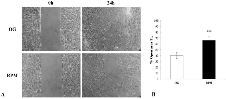
Figure 3.
Effects of simulated microgravity on wound healing in human dermal fibroblasts at 24 h. (A) Representative images show human fibroblasts exposed or unexposed to the RPM for 24 h at 0 and 24 h. Images were obtained by optical microscopy with 100× magnification. (B) Values are expressed as the percentage of open area, measured using ImageJ v 1.47 h software; the value of the open area at 0 h is 100%. Each column represents the mean value ± SD of four independent experiments with standard deviations represented by vertical bars. *** p < 0.001 RPM versus on-ground control (OG) by unpaired two-tailed t-test.

