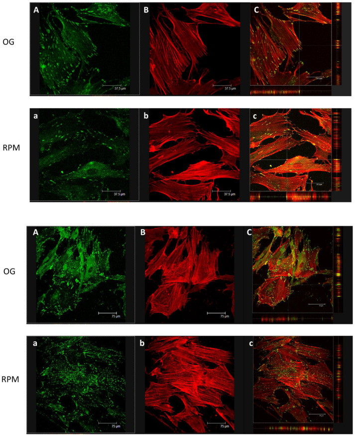
Figure 9.
(Upper panel) Representative confocal images of vinculin (green) and actin (red) in cells cultured under normal gravitational conditions (OG) and microgravity conditions (RPM) for 24 h. Fibroblasts in µG showed a remarkable reduction in vinculin distribution (a), particularly behind the cytoplasmic membrane and close to the cell’s free front, as well as a reduction of stress fibers (b) compared to OG condition (B). Vinculin (green) and actin (red) co-localized in fibroblasts cultured in normal gravity (C), especially at the level of protrusive structures (filopodia and pseudopodia). Little or no co-localization was observed at the level of membrane protrusion in an RPM-cultured cell (c) (scale bar 37.5 µm). (Lower panel) Representative confocal images of vinculin (green) and F-actin (red) in cells cultured in normal gravitational condition (OG) and microgravity conditions (RPM) for 48 h. Under RPM conditions, the organization of stress fibers was similar to that observed in cells maintained in normal gravitational (OG) conditions. (C,c) show Vinculin (green) and actin (red) co-localization (scale bar 75 µm).

