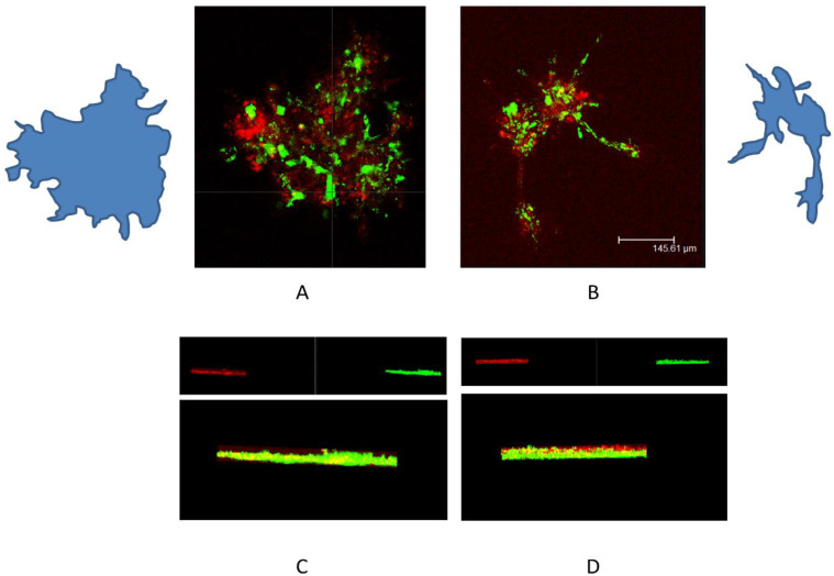
Figure 12.
Representative confocal images of fibroblast (green) and keratinocytes (red) co-cultured on Matrigel in normal gravitational (A) and microgravity conditions (B) for 24 h. (C,D) show the three-dimensional Leica software reconstructions. The confocal microscope analysis showed that in OG, the fibroblasts were arranged randomly with keratinocytes, while in the RPM, the fibroblasts and keratinocytes had distinct positions (scale bar 145.6 µm).

