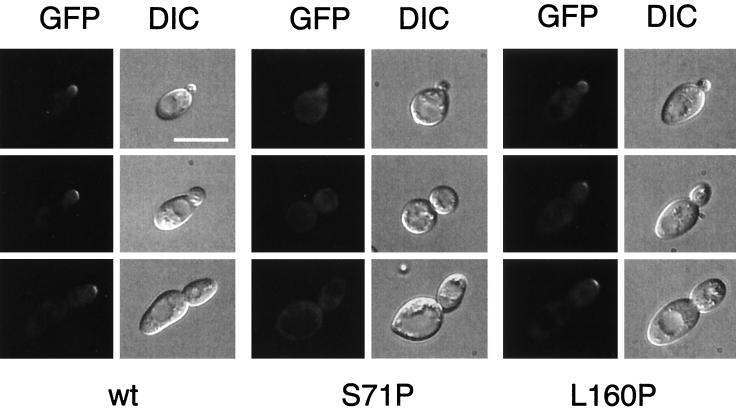
FIG. 6.
Subcellular localization of GFP-Ste20p in CDC42 pseudohyphal mutants. Shown are cells of diploid strains that express wild-type CDC42 (wt), CDC42S71P (S71P), or CDC42L160P (L160P) and harbor plasmid pME1760 encoding GFP-Ste20p under the control of the STE20 promoter. Living cells at different stages of the cell cycle were chosen for photography according to their bud size and were viewed by either fluorescence microscopy (GFP) or DIC microscopy. Identical results were obtained with centromere-based plasmid pME1759, although with markedly decreased fluorescence signals due to lower expression of GFP-Ste20p. Bar, 10 μm.

