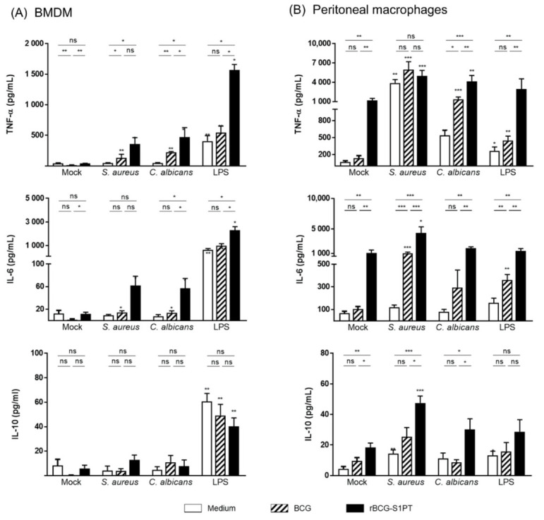
Figure 3.

Memory response of primed macrophages to heterologous challenges. BMDM (A) and peritoneal macrophages (B) from naïve mice were exposed in vitro to culture medium alone (unprimed, white bars), BCG (striped bars), or rBCG-S1PT (black bars) (MOI 0.1:1) for 24 h. Cells were then left to rest for 6 days and re-stimulated with medium alone (mock) or with S. aureus, C. albicans, or LPS (horizontal axis). The production of TNF-α (upper panels), IL-6 (middle panels), and IL-10 (lower panels) was measured after 24 h. Statistical analysis was performed via Mann–Whitney U test. * p < 0.05, ** p < 0.01, *** p < 0.001, ns = not significant. Bars represent mean ± SEM of 4–5 (for BMDM) and 5–8 (for peritoneal macrophages) replicate samples. Asterisks over the columns in S. aureus, C. albicans, and LPS refer to the comparison with the respective mock.
