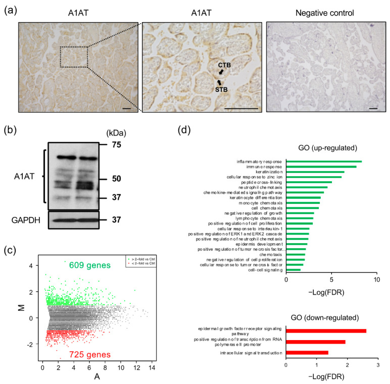
Figure 1.
Knockdown of A1AT upregulates the expression of inflammatory response-related genes in trophoblasts. (a) Expression of A1AT protein in human placentas. Immunostaining was performed using anti-A1AT antibody or normal rabbit IgG (negative control). The pictures display the chorionic villi. The magnified middle picture shows the rectangle in the left panel. Scale bar = 100 μm. CTB: cytotrophoblasts, STB: syncytiotrophoblast. (b) Lysates prepared from primary trophoblasts were subjected to immunoblotting, with GAPDH serving as the loading control. (c) MA plot showing the expression of transcripts identified by RNA-seq. The transcripts highlighted in red or green were more than 2-fold differentially expressed (p < 0.05). (d) Functional classification of differentially expressed genes (DEGs) by Gene Ontology analysis.

