Figure 4.
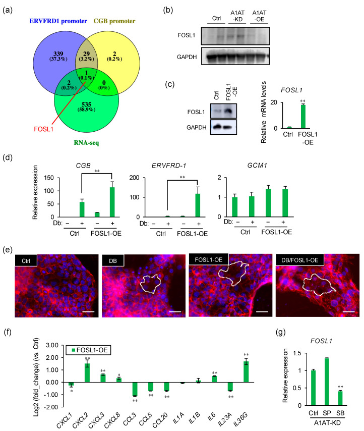
FOSL1 mediates A1AT knockdown-induced syncytialization of trophoblasts. (a) Venn diagram showing the numbers of upregulated DEGs and of genes encoding proteins that may be capable of binding to the ERVFDR-1 or CGB promoter. (b) Immunoblotting showing the protein levels of FOSL1 in lysates from A1AT-KD and A1AT-OE BeWo cells. GAPDH served as the loading control. (c) Expression of FOSL1 in FOSL1-OE BeWo cells by Immunoblotting (left) and qPCR (right). Results shown are the means ± SEMs of three independent experiments. ** p < 0.01. (d) Expression of syncytialization markers in FOSL1-OE BeWo cells treated with Db (0.5 μM) by qPCR. Values are means ± SEMs of three independent experiments. ** p < 0.01. (e) Visualization of syncytialization by immunostaining cells with anti-E-cadherin antibody (red) and DAPI (blue). Representative pictures are shown, with syncytialized cells marked with a stippled line. Scale bar = 50 μm. (f) Expression of mRNAs encoding inflammatory cytokines in FOSL1-OE BeWo cells. GAPDH was used as the loading control. Results are reported as the means ± SEMs of three independent experiments. * p < 0.05, ** p < 0.01. (g) Expression of mRNAs encoding inflammatory cytokines in A1AT-KD BeWo cells treated with SP600125 (SP, 20 μM) or SB203580 (SB, 20 μM) for 24 h. GAPDH was used as the loading control. Results are reported as the means ± SEMs of three independent experiments. ** p < 0.01.

