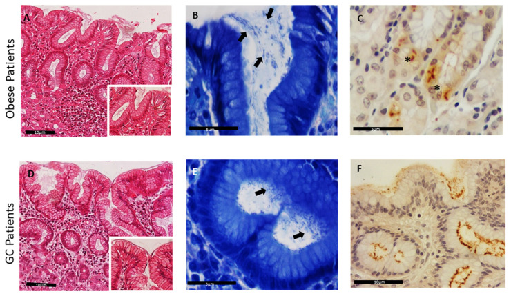
Figure 1.
Detection of Helicobacter species in human gastric mucosa sections. H&E stain of gastric sections from obese (OB) patients (A) and gastric cancer (GC) patients (D), 200× and 400× (inset). Modified-Giemsa stain, highlighting bacteria at the epithelium surface and gastric crypts (black arrows), in obese patients (B) and GC patients (E), magnification 600×. (C,F) Helicobacter spp. immunopositivity within the superficial mucus, in the lumen of gastric deep glands, 400×, and inside parietal cells, 600×, (*) in obese patients (C) and GC patients (F).

