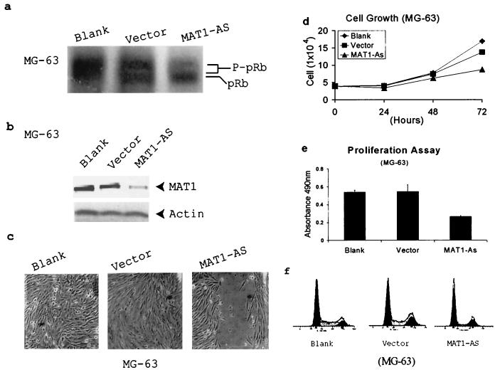
FIG. 1.
Deregulation of CAK leads to inhibition of pRb phosphorylation and G1 arrest in MAT1-AS-transduced MG-63 cells. (a) Western blotting depicts pRb phosphorylation. P-pRb, hyperphosphorylated form of pRb. (b) Western blotting detects MAT1 protein expression. Actin detection was performed on the same blot as the protein loading control. (c) Analyses of cell proliferation activation in an in vitro injured tissue. Transduced and nontransduced confluent MG-63 cells were scraped to release cells from contact inhibition. The time for closure of the wound track was assessed under a phase-contrast microscope. Blank and Vector cells show closure of the wound track at 72 h, while the wound track in MAT1-AS-transduced cells still was not closed at that time. The time for closure of the wound track in MAT1-AS-transduced cells was 192 h (data not shown). (d) Cell growth analysis. The same numbers of transduced and nontransduced MG-63 cells were plated; cell duplication was monitored by counting the cells up to 3 days before they reached confluence. The growth curves represent the mean and standard deviation from the cells of triplicate wells. (e) Cell proliferation assay. Transduced and nontransduced subconfluent MG-63 cells were incubated with MTS tetrazolium compound and quantified by measurement of the absorbance at 490 nm to determine the proportion of living cells in culture. The data represent the mean and standard deviation of triplicate wells. (f) Cell cycle analysis. The cell cycle profile in MAT1-AS-transduced cells showed 67% cells in the G0/G1 phase and 12% in the S phase, which is 34% more cells in the G0/G1 phase and 54% fewer cells in the S phase compared with controls.

