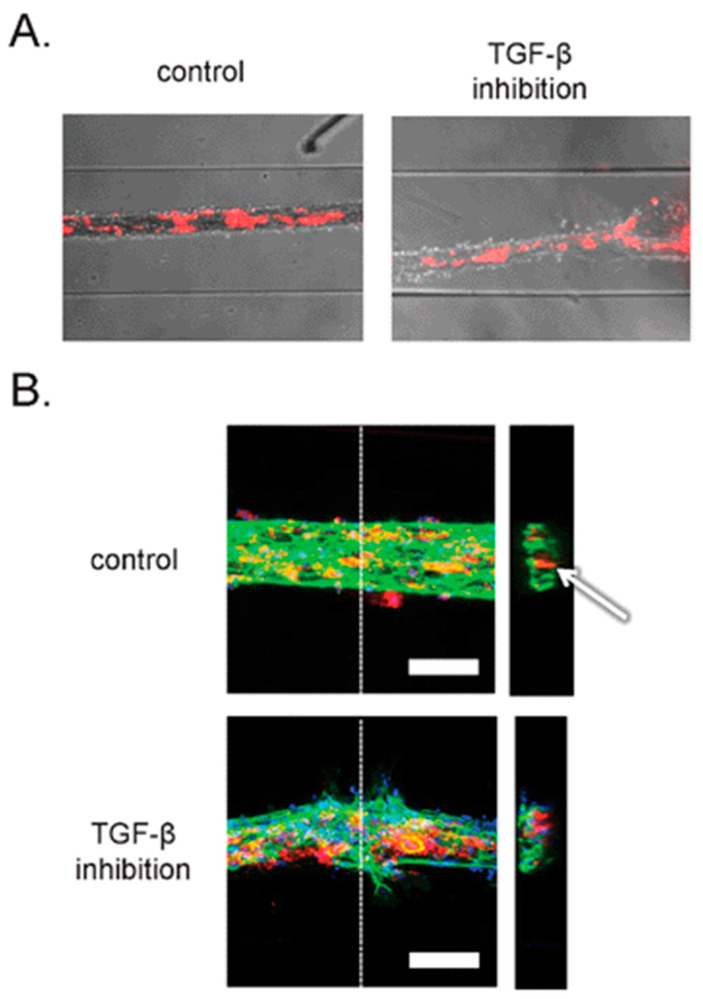
Figure 6.
(A) Brightfield images combined with epifluorescence images of an OOC platform. (B) Confocal microscopy images taken from the same OOC platform to obtain a higher level of detail of the 3D structures and interactions within this OOC device. Reproduced from [32] with permission, © 2013 Royal Society of Chemistry.

