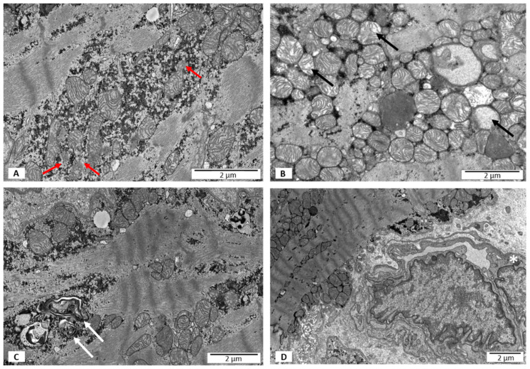
Figure 2.
Electron microscopy images. Ultrastructural features of heart biopsies from patients with EF < 50% and no virus detected. (A) A disrupted outer mitochondrial membrane marked with red arrows; (B) partial loss of mitochondrial cristae and swollen mitochondria marked with black arrows; (C) lysosomes and autophagous figures containing remnants of mitochondrial cristae marked with white arrows; (D) no features of necrosis; hypertrophic endothelium marked with an asterisk.

