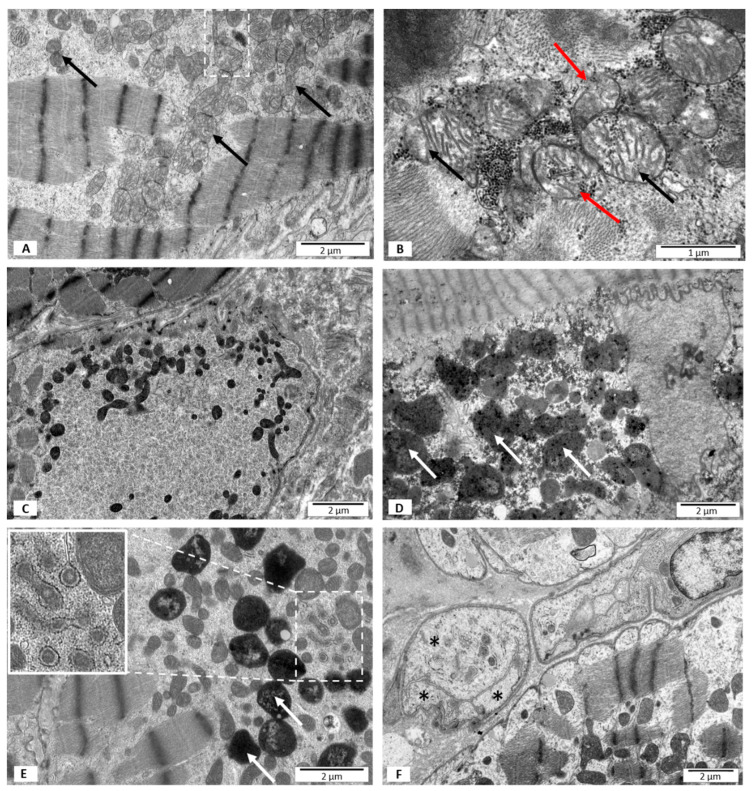
Figure 3.
Electron microscopy images. Ultrastructural features of heart biopsies from patients with EF < 50% and PB19 virus detected. (A) A blurred structure of mitochondrial membranes marked with black arrows; mitochondria showing morphological features of fission marked with frame; (B) a disrupted outer mitochondrial membrane marked with red arrows; glycogen granules in the mitochondrial matrix are visible. Mitochondria showing morphological signs of swelling marked with black arrows; (C) increased electron density of the mitochondria; (D,E) high mitophagy rate; mitophagosomes marked with white arrows; (E) sarcoplasmic reticulum with the amorphous material marked with frame and shown in the insert; (F) blood vessel with features of necrosis marked with asterisks.

