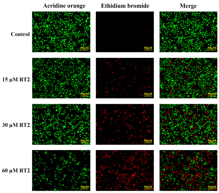
Figure 4.
Caco-2 colon cancer cell morphological changes. Caco-2 cells were treated with increasing concentrations of RT2 synthetic peptide (0, 15, 30, and 60 µM) for 24 h and then stained with dual acridine orange/ethidium bromide (AO/EB) fluorescent dyes. Under the fluorescence microscope (magnification 20×), the live cells and the early apoptotic cells are shown in green and yellow-green, respectively. The late apoptotic and necrotic cells appeared in orange and red, respectively. The merged images show co-distribution of live, apoptotic, and necrotic cells.

