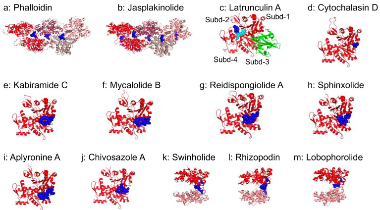
Figure 4.
Actin-binding small molecules: (a) Phalloidin (7BTI [54]). (b) Jasplakinolide (6T23 [99]). (c) Latrunculin A, (1ESV [100], Gelsolin domain 1 is shown in green); subdomains of actin are indicated as Subd-1~4. ATP is shown in cyan. (d) Cytochalasin D (3EKS [101]). (e) Kabiramide C (1QZ5 [102]). (f) Mycalolide B (6MGO [103]). (g) Reidispongiolide A (2ASM [104]). (h) Sphinxolide (2ASO ([104]). (i) Aplyronine A (1WUA [105]). (j) Chivosazole A (6QRI [106]). (k) Swinholide (1YXQ [107]). (l) Rhizopodin (2VYP [108]). (m) Lobophorolide (3M6G [109]). Actin-binding small molecules are shown in blue. Actin shown from pointed (left) to barbed (right) end with red.

