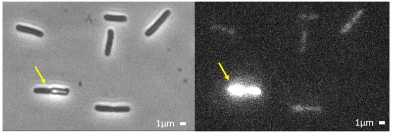
Figure 3.
Example of Phase contrast image (left) and of Sytox fluorescence (right) after 1 h flow of 4 μM L-AvBD103b. All bacteria are Sytox-labelled, generally very weakly. In a minority of cases, the high Sytox fluorescence testifies that both OM and CM were more strongly permeabilized (one strongly labelled bacteria in this example, yellow arrow).

