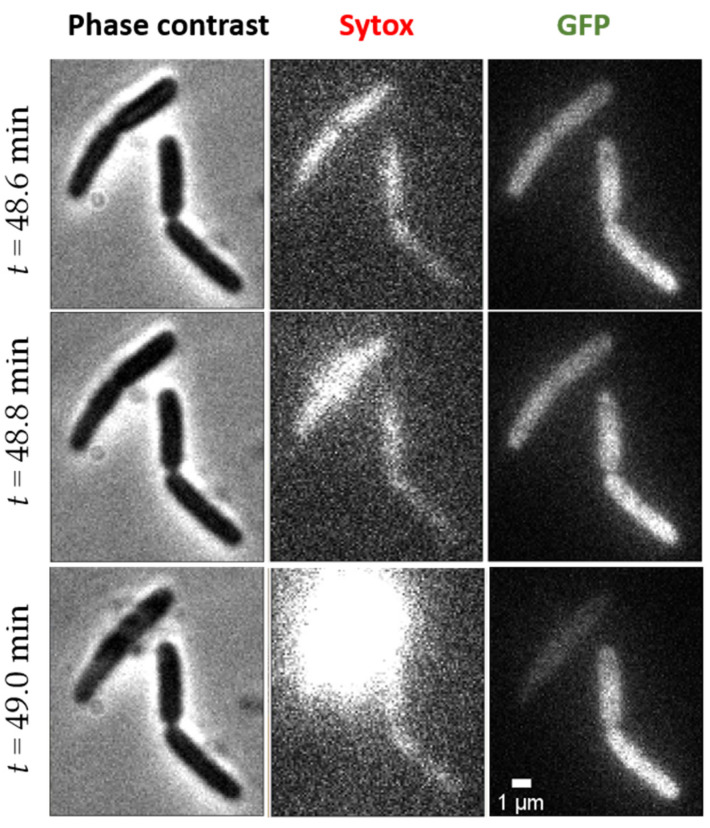
Figure 4.
Example of E. coli explosion occurring at time t = 49 min of exposure to 4 μM L-AvBD103b. Phase contrast (left), Sytox (middle), and GFP (right) images are represented at time 48.6 min (top, before explosion), at time 48.8 min (middle, very early sytox explosion), and at time 49 min (bottom, final state).

