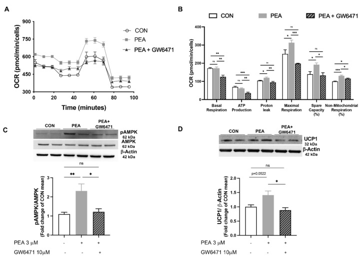
Figure 4.
PEA exerts its metabolic effects via PPAR-α activation in differentiated 3T3-L1 cells. (A) Mito stress assay is performed in differentiated adipocytes, in the presence or not of PEA (3 μM) and/or the PPAR-α antagonist GW6471 (10 μM), by the Seahorse analyzer XFe24; (B) key parameters of mitochondrial function are reported. Western blot analysis for (C) phospho-AMPK and (D) UCP1 are also displayed. Results are shown as mean ± SEM of three different sets of experiments for 3T3-L1, respectively. * p < 0.05, ** p < 0.01, *** p < 0.001 significantly differ from CON.

