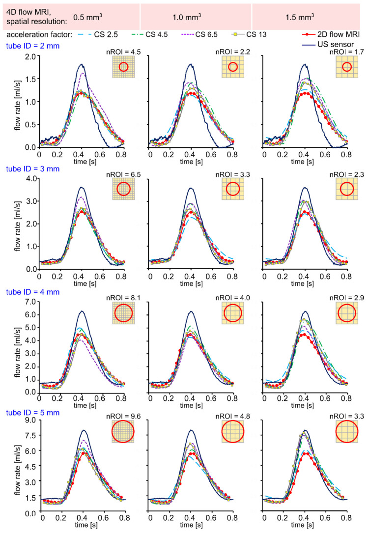
Figure 2.
Time-resolved flow rate curves measured with US sensor, 2D flow MRI, and 4D flow MRI in silicone tubes with an inner diameter (ID) of 2, 3, 4, and 5 mm, respectively. The flow rates were calculated in three ROI A-C, as shown in Figure 1, and then averaged. Qualitatively, the peak flow values were overestimated for lower resolution 4D flow MRI data in comparison to 2D flow MRI.

