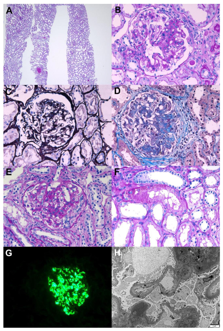
Figure 1.
Pathological findings of IgA nephropathy. Light microscopy: (A) In low magnification, no tubular atrophy or interstitial fibrosis was present (periodic acid–Schiff, ×40). (B) The glomerulus shows mesangial hypercellularity with increased mesangial matrix (periodic acid–Schiff, ×400). (C) The glomerulus shows endocapillary hypercellularity with focal loss of capillary penetrations (periodic acid methenamine silver, ×400). (D) The glomerulus shows segmental glomerulosclerosis (Masson’s trichrome, ×400). (E) The glomerulus shows cellular crescent with endocapillary hypercellularity (periodic acid–Schiff, ×400). (F) The picture shows protein reabsorption vacuoles in tubules, which is evidence of proteinuria (periodic acid–Schiff, ×400). Immunofluorescence: (G) Strong IgA mesangial positivity in the immunofluorescence stain (×400). Electron microscopy: (H) Numerous large electron-dense deposits in the mesangial areas with focal foot process effacement in the capillary surface were observed (×6000, 80 kv).

