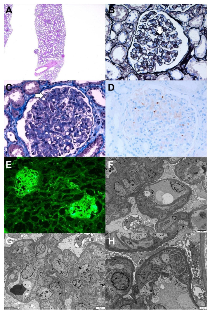
Figure 3.
Pathologic findings of chronic thrombotic microangiopathy. Light microscopy: (A) In low magnification, no tubular atrophy or interstitial fibrosis was present (hematoxylin and eosin, ×40). (B) The picture shows the duplication of the capillary basement membrane (arrow) (periodic acid methenamine silver, ×400). (C) The glomerular lumina were occluded due to the thickening of the glomerular capillary wall with a few fibrin thrombi (arrow) (Masson’s trichrome, ×400). (D) Immunohistochemical stain for CD61 (platelet glycoprotein GPIIIa) shows positivity against platelet thrombi (CD61, ×400). Immunofluorescence: (E) Strong positive staining for fibrinogen in the glomerulus was observed(×100). Electron microscopy: (F) Glomerular basement membrane duplication with cellular interpositions was observed (×3000, 80 kv). (G) Glomerular endothelial swelling and hypertrophy with occlusion of the lumens (glomerular endotheliosis) and diffuse foot process effacements along the capillary surfaces were observed (×3000, 80 kv). (H) Deposition of fibrinogen in the glomerular intracapillary area with entrapped cellular debris was noticed (×5000, 80 kv).

