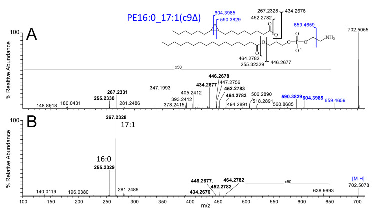
Figure 4.
Negative ionisation mode spectra obtained by (A) 213 nm UVPD-MS/MS and (B) CID-MS/MS for structural characterisation of the [M−H]− precursor ion of E. coli PE(33:1) observed at 733.5045 m/z in Figure 2 as predominantly containing PE16:0_17:1(c9Δ) under SGP conditions. The structure shown in the inset indicates the assigned bond cleavage sites for the major product ions. Product ions labelled in blue text are unique to UVPD.

