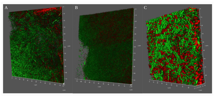
Figure 3.
Confocal microscopy images of B. haynesii CamB6 competing against oxygen stress in different media and forming different depth of pellicles (Live–death images are shown in the green- or red-false-colored fluorescence channels merged); (A) NB medium, (B) LB medium, and (C) MB medium after 96 h of optimum pellicle formation.

