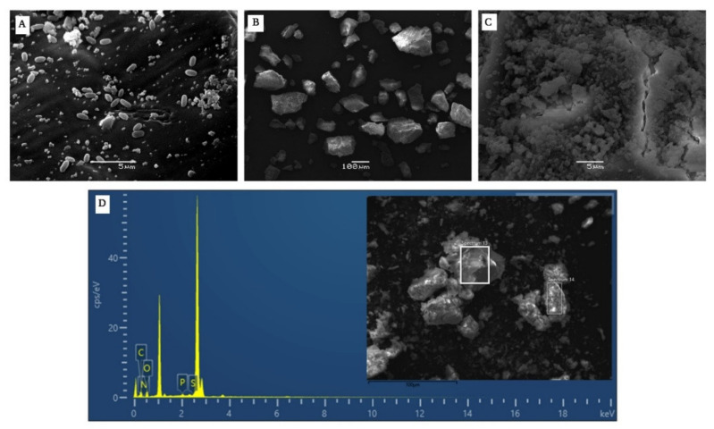
Figure 8.
SEM images showing (A) B. haynesii CamB6 cells producing melanin-like compound in MB at 96 h of incubation, (B) extracted melanin-like compound 1000×, and (C) at 5000× showing high density, compact, amorphous deposit with no definite pattern. (D) Elemental analysis of the melanin showing the presence of C, O, N, P, and S (inset pigment at 1000×).

