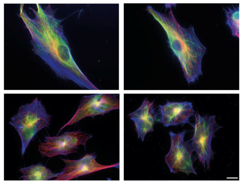
Figure 1.
Cytoskeletal organisation and shape of human fibroblasts for the different stages of metastatic transformation. Immunoflourescence staining followed by Epiflourescence 2D microscopy showing Bj primary fibroblast (top left), Bjhtert cells (top right), BjhtertSV40T cells (bottom left) and metastasising BjhtertSV40TRasV12 cells (bottom right), showing vimentin (green), tubulin (red) and F-actin (blue). Scale bar, 10 μm.

