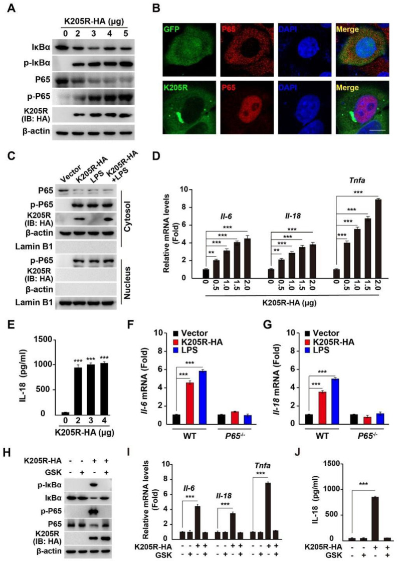
Figure 5.
ASFV K205R activates the NF-κB signaling pathway. (A) 3D4/21 cells were transfected with K205R-HA plasmid as indicated for 24 h. IκBα, p-IκBα, P65, p-P65, K205R-HA, and β-actin were assessed with immunoblotting analysis. (B) HeLa cells were transfected with empty vector or K205R-GFP plasmid for 24 h. The translocation of P65 into the nucleus was assessed with immunofluorescence analysis. Scale bar: 10 μm. (C) HeLa cells were transfected with K205R-HA and treated with LPS (1 mg/mL) as indicated for 24 h. P65 and p-P65 in the cytosol (indicated by β-actin) and nucleus (indicated by Lamin B1) were assessed with immunofluorescence analysis. (D) 3D4/21 cells were transfected with K205R-HA plasmid as indicated for 24 h. The mRNA levels of Il-6, Il-18, and Tnfa were assessed with qRT-PCR analysis. ** p < 0.01, *** p < 0.001. (E) 3D4/21 cells were transfected with K205R-HA plasmid as indicated for 24 h. IL-18 in the medium was quantified with ELISA. *** p < 0.001. (F,G) 3D4/21 WT and P65−/− cells were transfected with K205R-HA and treated with LPS (1 mg/mL) as indicated for 24 h. The mRNA levels of Il-6 (F) and Il-18 (G) were assessed with qRT-PCR analysis. *** p < 0.001. (H) 3D4/21 cells were transfected with K205R-HA plasmid and treated with GSK (10 μM) as indicated for 24 h. p-IκBα, IκBα, p-P65, P65, K205R-HA, and β-actin were assessed with immunoblotting analysis. (I) 3D4/21 cells were transfected with K205R-HA plasmid and treated with GSK (10 μM) as indicated for 24 h. The mRNA levels of Il-6, Il-18, and Tnfa were assessed with qRT-PCR analysis. *** p < 0.001. (J) 3D4/21 cells were transfected with K205R-HA plasmid and treated with GSK (10 μM) as indicated for 24 h. IL-18 in the medium was quantified with ELISA. *** p < 0.001.

