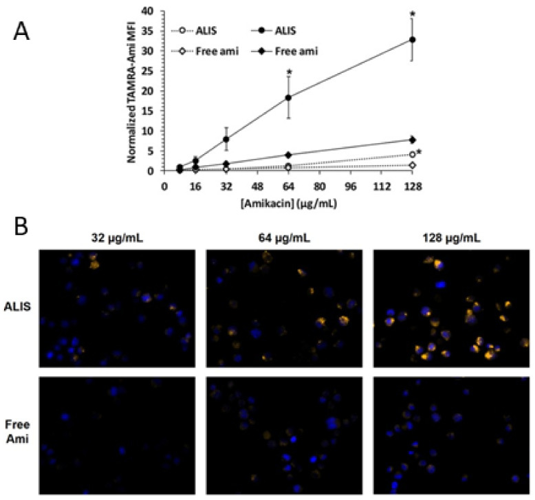
Figure 8.
Liposomal and free amikacin uptake into human macrophages. Macrophages were exposed to increasing concentrations of either ALIS or free amikacin (with addition of 0.44% tetramethyl rhodamine (TAMRA) conjugated amikacin) for 4 h (gray symbols) or 24 h (black symbols). (A) normalized mean fluorescence intensity (MFI) at each concentration averaged from three independent experiments. (B) Visualization of liposomal and free amikacin uptake in human macrophages by fluorescence microscopy. TAMRA fluorescence was visualized by a Zeiss Axio fluorescence microscope (400× magnification) using constant settings for all experimental conditions. Yellow: TAMRA amikacin; Blue: DAPI-stained DNA. * p < 0.05 vs. free amikacin at the same concentration and timepoint. Adapted from [149]. Frontiers, 2018. Furthermore, tissue and pulmonary macrophage exposures were compared in vivo after the 96 mg/kg amikacin dose delivered by ALIS nebulization versus the 100 mg/kg amikacin dose given intravenously [149]. In pulmonary macrophages, maximum amikacin concentration (Cmax) after the ALIS dose was nearly 1.0 μg/μg protein and the total macrophage exposure over 24 h (AUC) was 17.8 μg*h/μg protein, 274-fold higher than the exposure following amikacin injection. Similarly, the BAL fluid and lung tissue exposures were 69.5- and 42.7-fold higher, respectively, after inhalation dosing of ALIS compared to intravenous dosing. Simultaneously, the systemic (blood plasma) exposure was 5-fold lower for ALIS than for amikacin injection.

