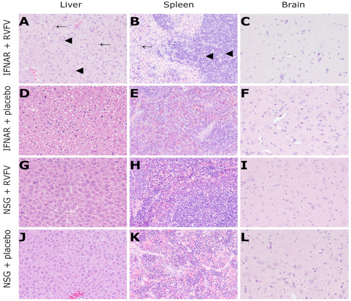
Figure 4.
Histology (HE staining, 200x magnification) of RVFV-infected IFNAR mice (3 dpi) and NSG mice (14 dpi): IFNAR mice exhibit severe, multifocal to coalescing hepatocellular necrosis (arrowheads, (A)) and apoptosis (arrows, (A)) as well as mild lymphocytolysis in the white pulp (arrowheads, (B)) and moderate, multifocal necrosis in the red pulp of the spleen (arrow, (B)). No lesions were observed in the brain (C) and placebo-infected control mice (D–F). Likewise, no lesions were present in the majority of RVFV-infected (G–I) or any placebo (J–L) NSG mice. RVFV: Rift Valley fever virus; dpi: days post infection; IFNAR: B6-IFNARtmAgt mice. NSG; NOD.Cg-Prkdcscid Il2rgtm1WjI/SzJ mice.

