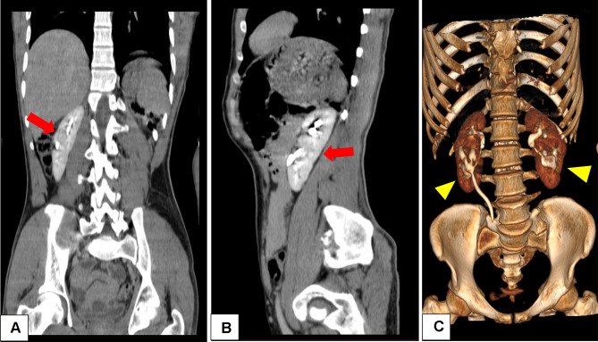
Figure 2.
Coronal and sagittal reconstruction of contrast enhanced CT scan showing the cleavage of fusion (arrows) between the native and supernumerary kidneys on the right side (A) and left side (B). Antero-lateral rotation of the supernumerary kidneys (arrow heads) and normal anatomic position of the native kidneys is also shown (C).

