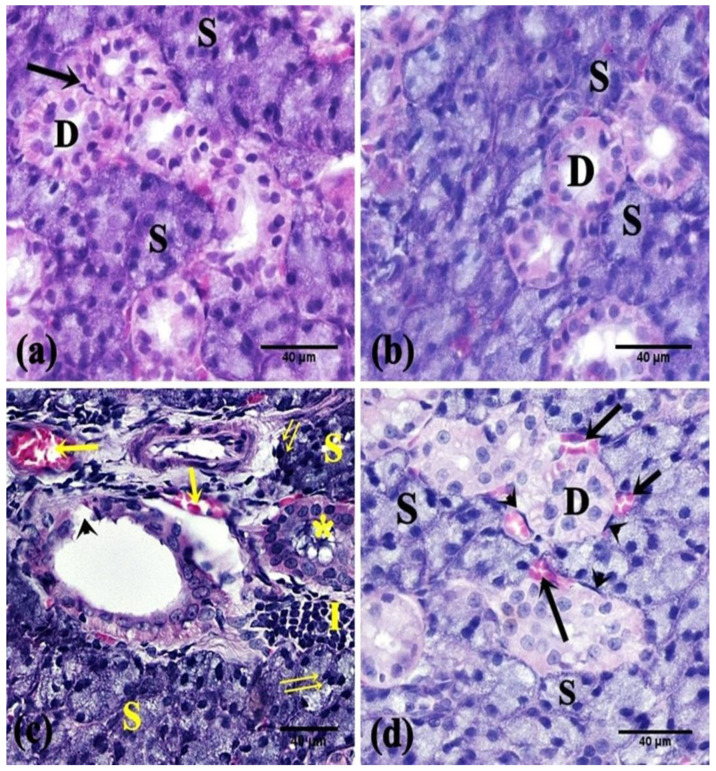
Figure 2.
Photomicrographs of the parotid salivary gland of the studied groups. (a,b) The control and FEB groups, respectively, showed intact parotid parenchyma; serous acini (S) and striated ducts (D). Note the myoepithelial cells (↑). (c) The 5-FU group showed parenchymal changes; coalesced serous acini (S), pyknotic nuclei (↑↑), cellular infiltration (I), interrupted lining of striated ducts (arrowhead), retained secretion (*), and dilated congested blood vessels (↑). (d) The FEB + 5-FU group showed normal parotid structure; serous acini (S), striated ducts (D), and myoepithelial cells (arrowhead) except for mildly congested blood vessels (↑). (H&E ×400; 8 rats/group).

