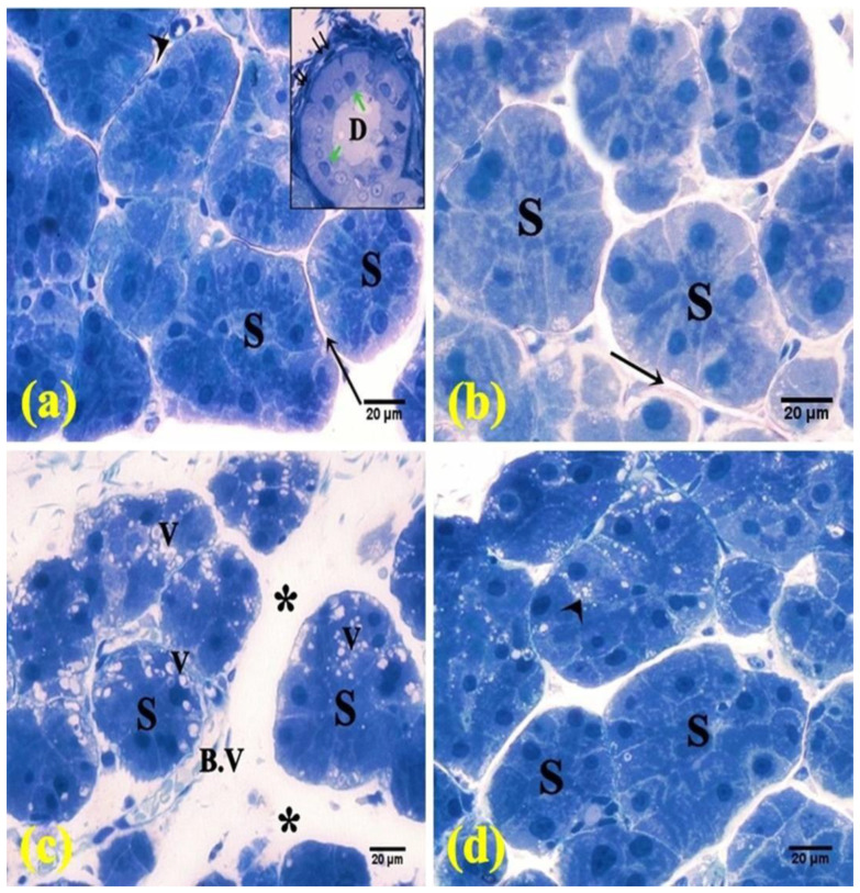
Figure 3.
Photomicrographs of semi-thin sections of the parotid salivary gland. (a,b) The control and FEB groups, respectively, showed intact parenchyma with packed serous acini (S), thin septa (↑), and blood vessel (arrowhead). Inset: striated duct (D) lined by low columnar cells (green arrow) and surrounded with myoepithelial cells (↑↑). (c) The 5-FU group showed serous acini (S) separated by wide spaces (*) and congested blood vessel (B.V). Notice the vacuolation (V). (d) The FEB + 5-FU group showed packed acini with few vacuolations (arrowhead). (Toluidine blue ×400; 8 rats/group).

