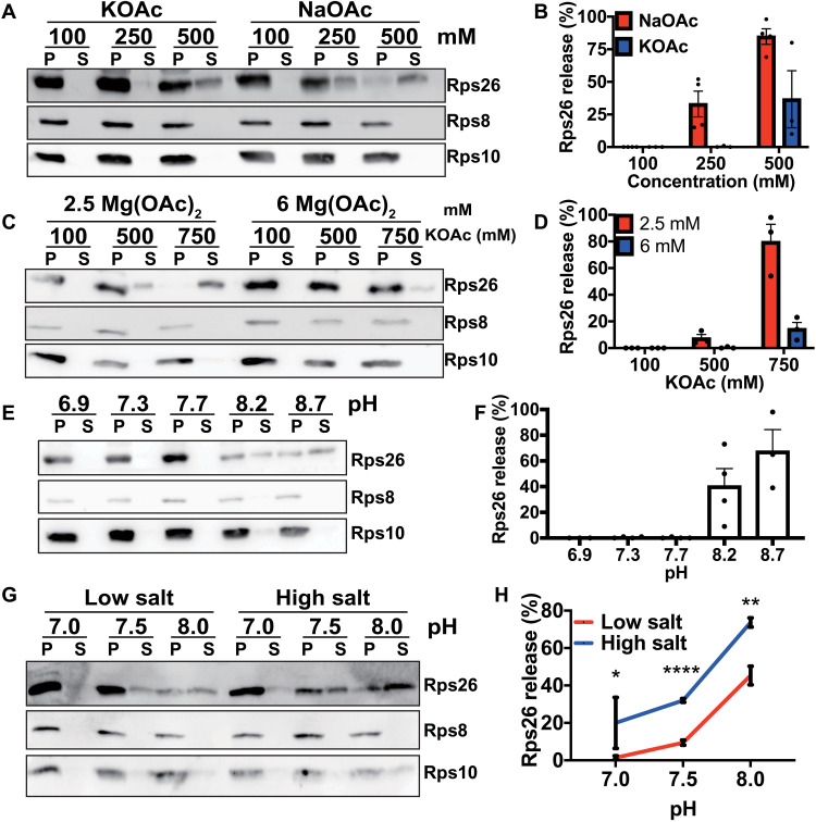
Fig. 5. Ribosomes directly sense ion and protein concentrations in vitro.
(A) Western blot analysis of pelleted (P) ribosomes and released proteins in the supernatant (S). Mature 40S subunits purified from yeast were incubated with recombinant Tsr2 at different concentrations of NaOAc or KOAc. (B) Quantification of Rps26 in (A) with three to four independent replicates for each condition. Error bars represent the SEM. (C) Effect of Mg+ in the release of Rps26. Mature 40S subunits were incubated with recombinant Tsr2 in either 2.5 or 6 mM MgOAc at different KOAc concentrations. (D) Quantification of Rps26 in (C) with three independent replicates for each condition. Error bars represent the SEM. (E) Release assay in 20 mM bis-tris propane at various pH values, 2.5 mM MgOAc and 100 mM KOAc. (F) Quantification of Rps26 in (E) with three to four independent replicates for each condition. Error bars represent the SEM. (G) Release of Rps26 under physiological conditions. Mature 40S subunits were incubated with recombinant Tsr2 in either low physiological salt conditions (20 mM NaCl, 200 mM KCl, and 2.5 mM MgOAc) or high physiological salt concentrations (150 mM NaCl, 150 mM KCl, and 2.5 mM MgOAc) with different pH values adjusted by bis-tris propane. (H) Quantification of results in (G) with three to nine independent replicates for each condition. Error bars represent the SEM. Significance was determined using an unpaired t test. *P < 0.05; **P < 0.01; ****P < 0.0001.

