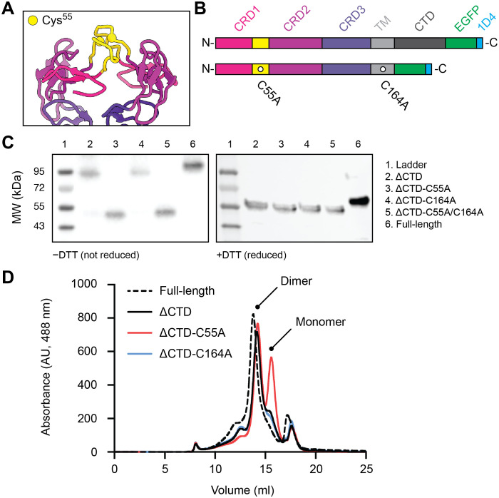
Fig. 2. The mGITR dimer interface is locked by an intersubunit disulfide bond.
(A) mGITR dimer interface with CRD1 (pink), CRD2 (maroon), and CRD3 (purple) and apical loops of CRD1 (yellow). Spheres on the apical loops mark Cys55. (B) Illustration of the mGITR protein with relevant domains. The full-length protein is shown at the top, and the mGITRΔCTD construct with C55A and C164A mutations is shown at the bottom. (C) Western blots of full-length mGITR and the four CTD deletion constructs. Nonreduced (−DTT) and reduced (+DTT) conditions are on the left and right, respectively. Each lane is numbered according to the sample it contains, and sample descriptions are provided in the legend. MW, molecular weight. (D) FSEC traces showing EGFP fluorescence of lysate from HEK293 cells expressing full-length mGITRegfp (dashed line), mGITRΔCTD-egfp (solid black line), mGITRΔCTD-egfp-C55A (red line), and mGITRΔCTD-egfp-C164A (blue line). AU, arbitrary units.

