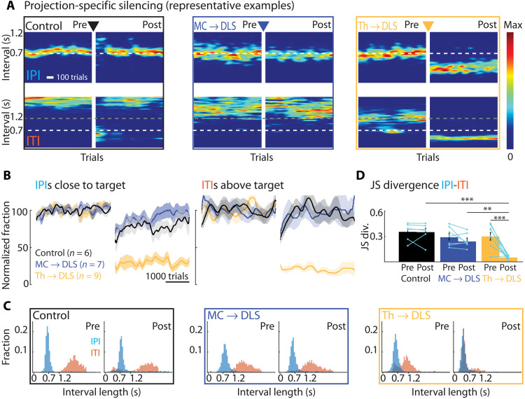
Fig. 4. Silencing DLS-projecting thalamus but not motor cortex neurons in expert animals disrupts performance of learned motor skills.
(A) Representative expert animals with silenced DLS-projecting neurons either in motor cortex (MC → DLS) or thalamus (Th → DLS) or control virus injections (see Results, Materials and Methods, and fig. S6). Shown are heatmaps of IPI and ITI probability distributions pre- and post-silencing (5 days post-surgery recovery between pre and post). (B) Population results for manipulations as in (A), normalized to performance before manipulation. Left: Fraction of trials with IPI close to target (700 ms ± 20%). Right: Fraction of trials with ITI above the threshold (1.2 s). Controls include animals expressing GFP in neurons either in motor cortex or thalamus projecting to DLS (n = 3 each) (see also fig. S6C). (C) Distributions of durations between lever-presses for animals shown in (A). Pre, last 2000 trials before silencing; post, first 2000 trials after silencing. (D) JS divergence as a measure of dissimilarity between IPI and ITI distributions for the same conditions as in (A). For statistical details, see table S4 and fig. S3D for further comparison of the manipulation effects. Bars show means, dots show individual animals, and error bars show SEM. **P < 0.01, ***P < 0.001.

