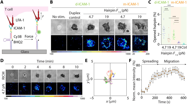
Fig. 2. Extracellular DNA-based LFA-1 tension probes.
(A) Design of covalently grafted ICAM-1 DNA-based tension probes. (B) RICM and tension images show that αCD3ε-treated naïve OT-1 cells spread and generated tension on probes with F1/2 of 4.7 or 19 pN after ~15 to 20 min of plating. Controls include nonprimed naïve OT-1 cells (no stim.) and αCD3ε-treated cells on control DNA probes that lack hairpin stem loop (duplex). d-ICAM-1, dimeric ICAM-1; m-ICAM-1, monomeric ICAM-1. (C) Plot showing average hairpin opening underneath the cell-substrate contact. Both monomeric (orange zone) and dimeric ICAM-1 (green zone) ligands were tested. n > 15 cells from at least two independent experiments. (D) Time-lapse RICM and tension images showing initial landing of αCD3ε-primed cell. (E) Displacement plot showing cell tracks of 10 randomly selected αCD3ε-primed cells on the tension probe substrates within 10 min. (F) Average LFA-1 tension evolution profile generated from 10 randomly selected αCD3ε-primed cells landing on the 4.7-pN tension probe substrates within the first 10 min. The mean intensities underneath T cells at each time point were normalized to the highest intensity within the sequence. ***P < 0.001; ****P < 0.0001. Scale bars, 5 μm. Probe density = ~1000 molecules/μm2.

