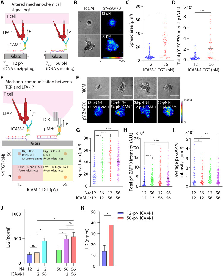
Fig. 4. Mechanochemical stabilization of LFA-1/ICAM-1 bonds potentiates TCR-triggered T cell activation and cytokine secretion.
(A) Schematic showing the design of TGT assays to cap maximal forces that can be transmitted to LFA-1/ICAM-1 bonds. (B) RICM and immunofluorescence images showing αCD3ε-primed naïve OT-1 cells spread on ICAM-1 TGT substrates after 1 hour of incubation. Cells were stained with Alexa Fluor 647–pY-ZAP70 antibody. (C and D) Quantification of spread area and pY-ZAP70 intensity of cells seeded on 12- or 56-pN ICAM-1 TGT substrates. n > 50 cells from three independent experiments. (E) Schematic showing the design of multifunctional surfaces presenting both ICAM-1 ligands (through the TGT) and immobilized agonist pMHC. (F) RICM and immunofluorescence images showing naïve OT-1 cells spread on these multiplexed TGTs after 1 hour of incubation. Cells were stained with Alexa Fluor 647–pY-ZAP70 antibody. (G to I) Quantification of spread area, total, and average pY-ZAP70 intensity of cells seeded on multiplexed TGT substrates. Dots represent cells pooled from three independent experiments. (J) IL-2 production by OT-1 (~1 × 10⁵ cells) after 6 hours of incubation on surfaces presenting N4 TGTs or N4 + ICAM-1 TGTs. (K) IL-2 production by αCD3ε-primed OT-1 (~1 × 10⁵ cells) after 24 hours of incubation on surfaces presenting only ICAM-1 TGTs. For (J) and (K), experiments were run using three batches of naïve cells. *P < 0.05; **P < 0.01; ****P < 0.001. Error bar represents means ± SEM. Scale bars, 5 μm. pMHC and ICAM-1 density are estimated to be ~400 to 500 molecules/μm2, respectively.

