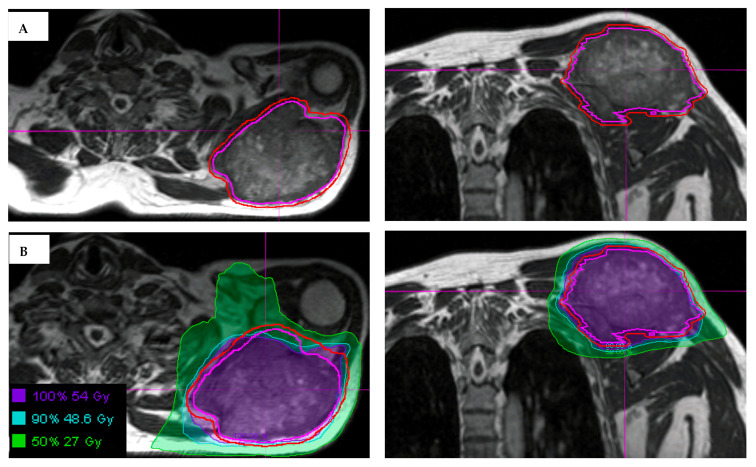
Figure 2.
(A) Visualization of an STS and normal tissues in the upper extremity using the 3D True Fast Imaging (TRUFI) sequence on MRI-guided linear accelerator. The gross tumor volume is outlined in magenta, with expansion to planning treatment volume (outlined in red) using small margins, in part due to MRI for daily set-up. (B) Hypofractionated radiation treatment plan for an upper extremity STS showing the use of MRI-guidance to delineate the tumor and the surrounding at risk areas.

