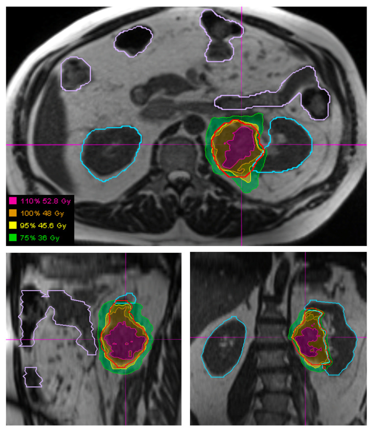
Figure 3.
Visualization of a retroperitoneal STS (outlined in red) and surrounding organs at risk, specifically bowel (outlined in light purple) and kidneys (outlined in light blue), using the 3D True Fast Imaging (TRUFI) sequence on a MRI-guided linear accelerator. Hypofractionated radiation plan showing the plan which was adapted each fraction to optimize bowel and kidney sparing.

