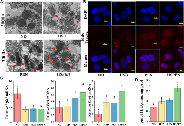
FIGURE 3.
HSD and antibiotic exposure induce mitochondrial dysfunction in the liver. (A) Swollen mitochondria and mitochondrial membrane rupture (red arrows) in the livers of mice as shown in typical TEM photomicrographs (×3000 magnification). (B) MitoTracker red was used to stain hepatocytes for 30 min and liver cells were analysed using confocal microscopy (scale bar: 10 μm). (C) The mRNA levels of the mitochondrial fusion marker Mfn1 and fission markers Drp1 and Fis1 in the liver tissue, normalized to levels in the liver tissue of the ND group. (D) Mitochondrial release of H2O2 in mouse liver. Data are shown as the mean ± SD; n = 8. Different letters were significantly different (p < 0.05).

