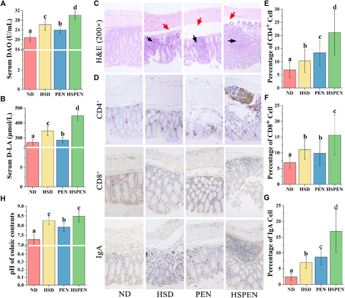
FIGURE 7.
Effects of HSD and antibiotic exposure on colonic function and morphology in mice. Levels of DAO (A) and D-LA (B) in serum, which signify the intestinal permeability of the ND-, HSD-, PEN- and HSPEN-treated mice. (C) Photomicrographs (×200 magnification) of the colon in the various groups of mice. Inflammatory cell osmosis and oedema in the submucosal layer are expressed respectively by black and red arrows. (D) Microscopic observation of CD4+, CD8+, and IgA lymphocytes on the colon epithelium (×200 magnification). Image Pro-Plus 6.0 software was employed to analyse the average numbers of CD4+ (E), CD8+ (F), and IgA (G) lymphocytes in the colon tissues and the (H) pH values of the colonic constituents of the groups. Data are expressed as the mean ± SD; n = 8. Different letters were significantly different (p < 0.05).

