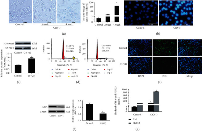
Figure 1.

Cr(VI)-induced premature senescence in L02 hepatocytes. (a) The L02 hepatocytes were treated with 10 nM Cr(VI) or PBS for 24 h twice a week for 2 or 4 weeks. The activity of SA-β-gal was assayed by β-Galactosidase Staining Kit (200x), and the percentage of senescent cells was showed in bar graph. After the L02 hepatocytes treated with Cr(VI) for 4 weeks, (b) the SAHF was examined by DAPI staining (400x), and (c) the protein level of H3K9me3 was determined by Western blot. (d) The cell cycle distribution was analyzed by flow cytometry analysis. (e) Edu was used to detect the inhibition of proliferation of L02 hepatocytes with Cr(VI) exposure (200x). (f) The expression of PCNA protein was assayed by Western blot. (g)The secretion of IL-6 and FGF23 was examined by ELISA kit. ImageJ software was used to analyze the relative levels of proteins normalized to the expression of GAPDH. All experiments were repeated at least 3 times and expressed as mean ± SD. ∗p < 0.05, compared with control. For the sake of clarity, the same control GAPDH was applied to compare with all experimentally relevant proteins with the same exposure time detected on the same SDS-PAGE gel unless otherwise stated.
