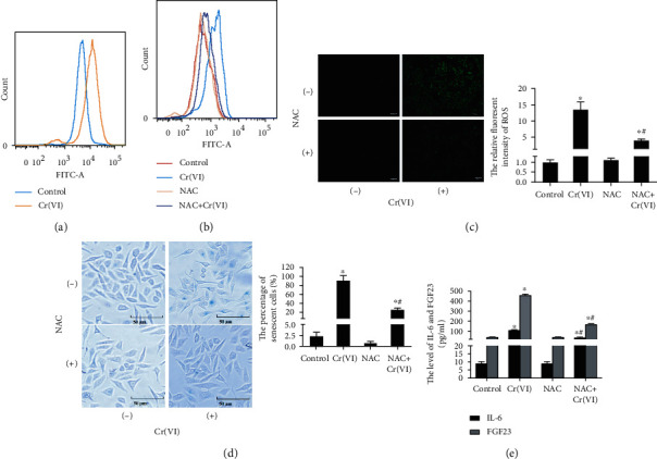
Figure 2.

Cr(VI)-induced premature senescence via intracellular ROS formation. (a) The L02 cells were treated with Cr(VI) for 4 weeks and loaded with DCFH-DA. The mean DCF fluorescence was measured by flow cytometry. The cells were incubated for 1 h in the presence or absence of NAC (10 mM) prior to Cr(VI) exposure. The mean DCF fluorescent intensity was assayed by (b) flow cytometry and (c) fluorescent microscopy (200x), and ImageJ software was used to analyze the relative fluorescent intensity showed in bar graph. (d) After same treatment, the SA-β-gal activity was determined by β-Galactosidase Staining Kit (200x), and the percentage of senescent cells was showed in bar graph. (e) ELISA was used to detected the levels of IL-6 and FGF23. All experiments were repeated at least 3 times and expressed as mean ± SD. ∗p < 0.05, compared with control.
