
Figure 6.
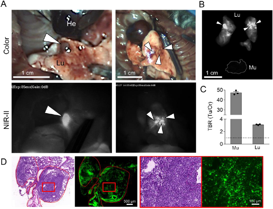
Orthotopic lung tumor targeting. A) 50 nmol of SH1 was injected into the mouse model of orthotopic lung cancer tumors 48 h prior to imaging. White arrowheads indicate lung tumors. (excitation = 808 nm; power density = 30 mW cm−2; exposure time = 25 ms; optical filter = 1,070 nm LP) B) NIR images and C) tumor-to-background signal ratios of dissected tumors compared with muscle and lung (n = 3, mean ± s.e.m.). D) Postoperative histopathological examination; H&E staining images and NIR fluorescence microscopic images.