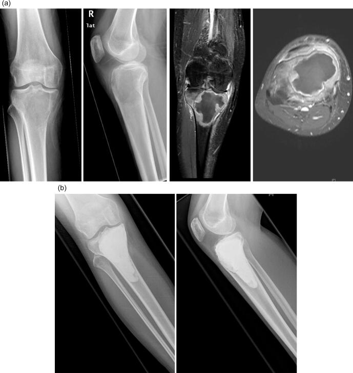
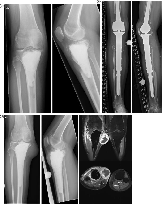
Fig. 3.
a-e: case of a 39-year-old female patient with GCTB of the proximal tibia: 3a: radiographs (left and middle) and MRI-scan (middle and right) of the proximal tibia showing an osteolysis; 3b: postoperative radiograph after intralesional curettage and defect reconstruction with bone cement; 3c radiographs of first local recurrence 18 months after surgery; 3d: radiographs (left and middle) and MRI-scan (middle and right) of second local recurrence 9 months after second curettage; 3e: radiographs of a modular tumor endoprosthesis which had to be implanted due to massive bone defect.


