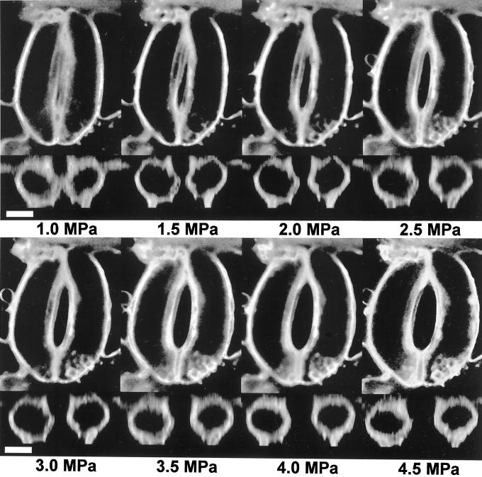
Figure 2.
Composite figure showing medial paradermal (x-y plane) and transdermal (x-z plane) images of guard cells as affected by turgor pressure. The pressure probe was inserted through the top of the left cell and into the right cell, and this caused both cells to fill with oil (see “Materials and Methods” for details). Scale bar = 10 μm. Movies showing three-dimensional reconstructions of guard cells can be found at http://bioweb.usu.edu/kmott or www.plantphysiol.org.

