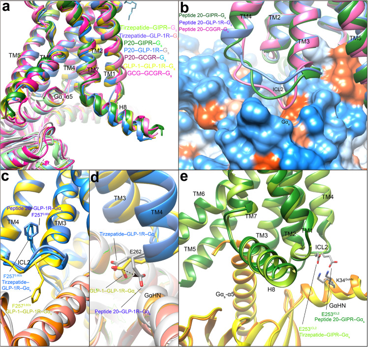
Fig. 6. G protein coupling of multi-targeting agonist-bound GIPR, GLP-1R and GCGR.
a Comparison of G protein coupling among GIPR, GLP-1R and GCGR4, 25, 27. The Gαs α5-helix of the Gαs Ras-like domain inserts into an intracellular crevice of receptor’s TMD. The receptors and G proteins are colored as the labels. b Comparison of ICL2 conformation in the peptide 20-bound GIPR, GCGR and GLP-1R. c Comparison of F2573.60b conformation in the GLP-1R bound by GLP-1, tirzepatide and peptide 20. d Comparison of E262ICL2 conformation in the GLP-1R bound by GLP-1, tirzepatide and peptide 20. e Comparison of E253ICL2 conformation in the GIPR bound by tirzepatide and peptide 20. Residues involved in interactions are shown as sticks. Polar interactions are shown as black dashed lines.

