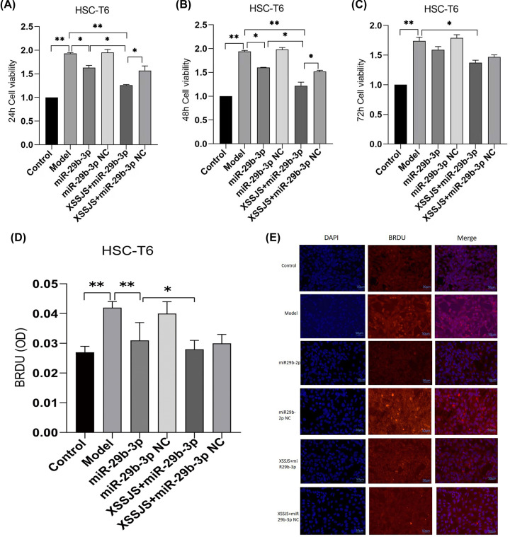
Figure 3. The cell proliferation was tested by using CCK8 and BrdU IF.
(A–C) The CCK8 proliferation assays (taken at 24, 48, 72 h, respectively) showed that XSSJS+miR-29b-3p better prevented the proliferation of HSC induced by TGF-β than XSSJS and miR-29b-3p at 24 and 48 h. There is little difference between their cell viability at 24 and 48 h, while the viability at 72 h is less effective than the viability at 24 or 48 h. (D) BrdU images showing that intervention with XSSJS+miR-29b-3p could more significantly decrease the number of BrdU-positive cells than XSSJS or miR-29b-3p administration. Bars = 50 μm. (E) Graphs of BrdU-positive ratios. All the experiments were repeated three times and represented as the means ± SEM (x ± s, n=3, *P<0.05, **P<0.01).

