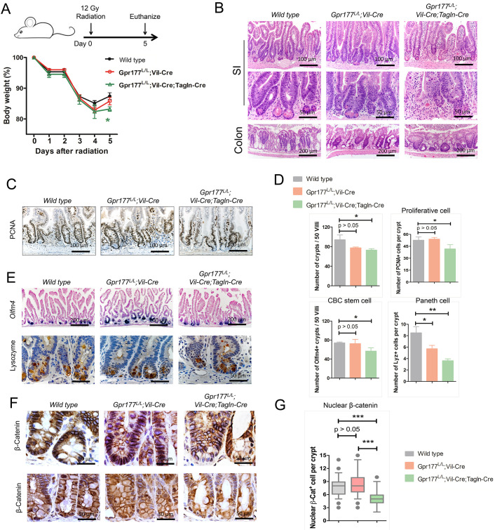
Fig. 5.
Blocking Wnts from epithelium and Tagln+ cells modestly affected irradiation-induced regeneration. (A) Schematic diagram and body-weight analysis of mice exposed to 12 Gy total body irradiation. The Gpr177L/L;Vil-Cre;Tagln-Cre mice showed a modestly significant body-weight reduction at day 5 after irradiation. n=6 mice per genotype. (B,C,E) Post-irradiated Gpr177L/L;Vil-Cre;Tagln-Cre mice had fewer regenerative crypts (B), PCNA+ proliferating crypt cells (C), Olfm4+ cells in the crypt (E) and Lyz1+ cells in the crypt (E). Gpr177L/L;Vil-Cre mice showed modestly reduced Lyz1+ cells. (D) Number of crypts, PCNA+ cells, Olfm4+ crypts and Lyz1+ cells were quantified for wild-type, Gpr177L/L;Vil-Cre and Gpr177L/L;Vil-Cre;Tagln-Cre mice intestines shown by representative images. n=3 mice per genotype. (F,G) Gpr177L/L;Vil-Cre;Acta2-CreER mice showed reduced number of nuclear β-catenin+ cells in regenerating crypts of distinct small intestinal regions (n=3 mice per genotype). Boxes in G represent 2nd and 3rd quartiles, and bottom and top whiskers represent 10% and 90% of the data. Outliers are shown as dots. Data are mean±s.e.m. from at least three independent experiments. ***P<0.001.

